Automating Particulate Inspection: More Than Meets the Eye
The most commonly used instrument for the detection of unwanted particles in drug manufacturing is the human eye, and manufacturers have established quality protocols and procedures governing how product is to be inspected, how long a particular inspector can be perform his/her task, and how often the inspection process is challenged. Whether relying on the naked eye or magnification under carefully controlled illumination, human inspectors are effective when the product type and packaging change often, the production throughput is low (with adequate inspection time) and the product requires manipulation to inspect.
However, market demands are continuing to push for higher throughput at increasing levels of product quality, meaning less time for operators to inspect and higher false reject or accept rates unless more staff are brought on board. The result is that industry is exploring automated alternatives to visible particulate detection.
Unfortunately, an off-the-shelf “all kinds of particulate in any kind of package” solution does not exist. Automated techniques vary depending on particulate, product and container properties. Some combinations of particle, product and container lend themselves to inexpensive technology. Other combinations require a more exotic approach, where the cost of the solution needs to be weighed more carefully against the cost of the problem.
This article will focus on automated techniques for detecting visible particles and help readers better understand the degree of complexity associated with various forms of particulate inspection.
Particulate Particulars
At its most basic, particulate contamination are solid objects in your container that you do not wish to be there. These can be foreign particles that have entered from the outside environment or they could be solids that have formed out of solution.
Particulate can be stable in solution, such as container fragments or they can react with the container contents, such as iron filings in a peroxide solution. Obviously, particulate that reacts in solution is a very serious issue. However, seemingly harmless particulate could cause unexpected end user quality issues as well.
For instance, a container or delivery mechanism may have a barrier filter to prevent particulate matter from escaping the container. However, particulate could block the filter and impede flow over time. In the case where no barrier filter is present, such as with injectable products, small particulate could enter the patient with potentially fatal results.
In the regulatory world, there is a distinction between visible and non-visible particulate matter. Visible particulate is loosely defined as any particulate that can be detected with the unaided eye. Typically, visible objects are defined as objects that are 0.1 mm or larger, which are about the average diameter of a human hair although some have pegged the limits of human detection as low as 50 um.
There is precious little in terms of regulatory guidance when it comes to visible particulate matter. The most referenced guidance comes from the USP, most notably in General Chapter—Injections <1> and in <788> (although <788> generally deals with sub-visible particles).
USP <1> states that each preparation is “essentially free” from visible particles and that every container that shows evidence of visible particles shall be rejected. USP <788> also reiterates the “essentially free” requirement. As designers of processes, we are left to interpret the meaning of “essentially free” along with a vague definition of how big a visible particle actually is.
In the case of sub-visible detection, there are more specific guidance’s available for process design. Specifically, ICH Guidance for Industry Q4B—Annex 3 Test for Particulate Contamination: Subvisible Particles General Chapter (January 2009), which references USP <788> among others. Particulate sizes, counts per volume as well as automated and manual techniques are outlined. Generally, tests for sub-visible particles are destructive and require samples to be taken from a lot for qualification. Sub-visible particles fall beyond human acuity and require microscopy or similar techniques for detection.
How Difficult to Detect?
Visible particulate inspection is generally done while the product is in its container, be it in solution, lyophilized cake, powder or ointment. In an ideal world the container would be clear, uniform and free from defects, but this is generally not the case. Containers can be coloured, frosted and opaque.
Also, visual inspection of solid product is limited to the surfaces of the product that are exposed. The product would also have to be inspected from a variety of angles to ensure each surface was examined. The interior of a solid product cannot be inspected unless the inspection system uses a probe that can penetrate the solid.
The ease with which a particle can be detected depends on the following factors:
- The size of the particle: Of course, the bigger the particle, the easier it will be to detect. Particles on the order of 100 um in diameter are challenging, while particles 1 mm or larger are much simpler.
- The contrast of the particle: A black particle on a white background is easier to detect than a clear particle in a clear solution.
- The density of the particle: A particle that is much denser than its surroundings or its container will easier to detect using more exotic inspection methods.
- The transparency of the container: It stands to reason that dark, cloudy or opaque containers will offer less visibility to the particulates inside. Amber vials are a more difficult container for particulate inspection than clear containers.
- The uniformity of the container: non uniform surfaces on the container such as variations in wall thickness can cause havoc on illumination methods due to refraction of light at the surface. These surfaces include the curved areas of a glass vial or the side walls of a moulded plastic bottle.
To put the relative difficulty of detection into perspective, a 1-mm diameter black particle sitting on top of white powder in a clear vial is generally easily to detect with readily available equipment. A 1-mm partially transparent particle floating in an IV bag can be detected with repeatability, but the inspection would require specialized equipment and software algorithms.
Clear Solution in Clear Container
A good example of clear particulate in a clear container can be found in IV bag inspection. Inspection typically occurs after the bags have been labelled, filled and sealed. Inspection is achieved manually and the inspection process can include other quality checks in addition to particulate inspection. Operators may have on the order of seconds to inspect, less than a second for accelerated line rates. Depending on the procedure, illumination may not be much more than ambient. Also, after filling the solution may contain many bubbles, which could easily hide particulate such as ball bearings (which may look like bubbles) or clear plastic fragments. If the bag already has a printed label on it there is the additional problem of the labelling obscuring the bag contents.
The most common approach to automating the inspection for particulate in the above situation is to agitate the bag and the image the contents of the bag over time. The amount of time is usually limited by the line production rate. The imaging system generally consists of a machine vision camera, illumination (in this case backlighting) and a vision processor to analyze the images.
In the past, it was common for the vision camera and the vision processor to be separate. This was because the processing power required for analysis called for better than average computing power. However, advances in processing capability has made it possible to achieve the task using “smart camera” technology whereby the vision camera and the vision processor are in a single package.
It should also be noted that the method used for agitation will vary, depending the particulate type and size to be located. In general, a system will have to remove the bag from is carrier (usually a conveyor), fixture the bag such that it can be agitated, held in place for imaging, and then placed back on the carrier (or discarded as a reject). Depending on the nature of the production line, such a system will likely require custom engineering and fabrication to ensure proper functionality with the product and production setting.
Once the images have been acquired, they are then analyzed in sequence for image-to-image differences. If the IV bag is held firm during imaging, the differences can be interpreted as objects moving inside the bag such as gas bubbles and particulate. In the case of larger, denser particulate, detection is achieved by filtering out the gas bubbles from the analysis, as they will rise up while the particulate sinks.
Figure 1 shows images of 1-mm and 2-mm clear plastic particles being detected in a 1-L IV bag using motion tracking. The agitation was achieved by rocking the bag from side to side a number of cycles and then imaging immediately after the final cycle. It should also be noted that packaging plays a role in an automated system’s detection ability. If the interior surface of the container is irregular or contains sharp transitions, there will be pockets in which particulate can get stuck. In some cases, only vigorous agitation can dislodge the particle from its crevasse, creating excessive gas bubbles in the process.
If the goal is to find smaller, less dense particulate such as hairs, sand, or plastic smaller than 1 mm in diameter, a more careful approach to agitation must be used to remove gas bubbles from the field of view. This can be done by agitating the bag through spinning, with careful attention to acceleration and deceleration rates.
Clear Solution and Opaque Container
Despite a manufacturer’s best efforts, particulate can get into a container in a sterile filling environment. If the bottle is clear, the methods for inspecting for particulate would likely follow an agitation and image methodology. If, however, the solution is in an opaque container (due to light sensitivity or marketing requirements), traditional imaging is not possible. Instead, a technique that utilizes radiation that does penetrate is required.
An effective and increasingly more common system for detecting non-ferrous particles in opaque objects is x-ray imaging. Advances in emitters and detectors have allowed x-Ray technology to grow into more industrial settings, and there are a number of commercially available x-ray systems for industry. Off-the-shelf x-ray components, such as high-resolution cameras, are also widely available and make it possible for custom applications to be developed where they were not available in the past.
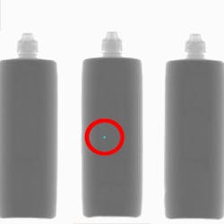
In the context of particulate inspection, x-ray systems are essentially density detectors. Differences in density through an object will show up as light or dark patches on a resultant image. The density difference between the particle and the surrounding area determines the degree of contrast that will be observed. With this in mind, the limitations of the approach become clear. If the density of the particle is similar to the surroundings, the contrast will be low, as will be the particle detectability. Figure 2 shows an image of a 0.8-mm stainless steel particle detected in a 120-ml opaque container using an off-the-shelf x-ray inspection system.
Opaque Solution and Clear Container
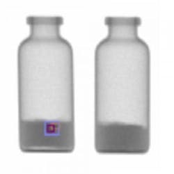
Another good application of x-ray technology is to look for particulate embedded in the interior of a powder or lyophilized product. Embedded glass is a particularly challenging problem, as it can enter the container at multiple stages of the production cycle. In fact, glass is difficult to detect even when it is not embedded in the product. Nevertheless, the density difference between the glass and surrounding material creates enough contrast under x-ray to been seen by an automated system. Figure 3 illustrates glass in powder under x-ray imaging.
Testing and Calibration
Generally, testing for particulate detection systems is achieved using calibrated particle size standards. These micro spheres can be made out of a variety of materials including polystyrene and stainless steel. Testing of an automated particulate detection device is achieved by spiking known good product with the spheres under production conditions. Spheres can be clear or opaque and range in size from millimetres down to fractions of a micrometer. When selecting the correct microsphere, it is important to bear in mind the size, opacity, and density of particulate for which the inspection system has been designed. If an appropriate microsphere is not available, then an alternate solution would be to collect or manufacture representative defects considered to be marginal, typical or gross defects. If this method is chosen, ensure that neither defect nor the solution it is suspended in change over time.
About the Author
Josh Capogna is currently a Manager at McRae Integration (Toronto), and is Founder and Senior Consultant at ODD Solutions.


